
Abstract:
Partial secondary edentulism, the loss of teeth in a partially dentate arch, presents unique challenges for implant-supported rehabilitation. While single-tooth implants are increasingly common, often a more complex treatment plan involving multiple implants is required. Two-stage implant placement remains a cornerstone technique for these cases, offering predictability and flexibility. This review comprehensively examines the principles underpinning two-stage implant placement in partial secondary edentulism, covering patient selection, comprehensive diagnostics, surgical protocols (including site preparation, implant selection, and placement techniques), timing of restoration, prosthetic considerations, and potential complications. We will explore the importance of bone grafting, soft tissue management, and the role of advanced technologies in optimizing treatment outcomes. This review aims to provide a practical guide for clinicians involved in the restoration of partially edentate arches with implant-supported solutions.
1. Introduction
Partial secondary edentulism is a prevalent condition affecting a significant portion of the adult population. The consequences extend beyond aesthetics and function, impacting chewing efficiency, speech, and overall quality of life. Traditionally, treatment options included removable partial dentures (RPDs), fixed partial dentures (FPDs), and more recently, implant-supported restorations. While RPDs and FPDs remain viable options, implant-supported solutions are increasingly favored due to their superior retention, stability, and preservation of adjacent tooth structure.
Two-stage implant placement, a well-established surgical protocol, allows for osseointegration followed by a secondary surgical exposure for prosthetic access. This approach provides flexibility in restoration design and timing, making it particularly suitable for complex cases involving multiple missing teeth and varying bone quality/quantity. This review will focus on the core principles guiding successful two-stage implant placement in the context of partial secondary edentulism, covering all aspects from initial assessment to long-term maintenance.
2. Patient Selection & Comprehensive Diagnostics
Successful implant treatment begins with meticulous patient selection. A thorough medical history is crucial to identify systemic conditions (e.g., uncontrolled diabetes, smoking, osteoporosis) that could compromise osseointegration or healing. Parafunctional habits (e.g., bruxism, clenching) must be assessed as they influence implant loading protocols and prosthetic design.
Comprehensive diagnostics are paramount and include:
- Clinical Examination: Assessing remaining teeth for periodontal health, mobility, and occlusion. Evaluating soft tissue contours, frenum positions, and mucogingival junctions.
- Radiographic Evaluation: Cone-Beam Computed Tomography (CBCT) is the gold standard for evaluating bone volume and density, nerve and sinus locations, and anatomical variations. Panoramic radiographs provide a broader overview, while periapical radiographs assess localized bone conditions.
- Digital Impressions & 3D Imaging: Facilitates accurate model creation for treatment planning and prosthetic fabrication.
- Photographic Documentation: Essential for aesthetic evaluation and documentation of pre-treatment conditions.
- Occlusal Analysis: Identifies existing occlusal imbalances and guides prosthetic design to ensure harmonious function.
- Study Models: Used to visualize the arch form and assess interocclusal space for implant placement.
3. Surgical Protocol: Site Preparation
Site preparation is critical for achieving primary stability and optimal bone-to-implant contact. This involves:
- Flap Design: A full-thickness mucoperiosteal flap is typically elevated to expose the underlying bone. Flap design should prioritize soft tissue preservation and adequate access for implant placement. Various flap designs exist (e.g., crestal, modified crestal, envelope flap) depending on the clinical situation.
- Osteotomy Preparation: Guided implant surgery, utilizing surgical guides fabricated from CBCT data, is increasingly common. This allows for precise implant placement in the planned position and angle. Osteotomy preparation should be performed in a stepwise manner, starting with a pilot drill and gradually increasing in diameter. Adequate cooling with sterile saline is essential to prevent bone necrosis.
- Bone Grafting: Often necessary in cases of insufficient bone volume. Various bone grafting materials exist, including autografts (e.g., iliac crest bone), allografts (e.g., freeze-dried bone allograft), and synthetic bone substitutes (e.g., hydroxyapatite). The choice of grafting material depends on the defect size and patient-specific factors. Guided Bone Regeneration (GBR) techniques, utilizing membranes to contain the graft material, can enhance bone regeneration.
- Sinus Lift: When implant placement in the posterior maxilla is planned, a sinus lift may be necessary to augment bone height. This involves elevating the Schneiderian membrane and grafting the resulting space with bone material.
4. Implant Selection
Implant selection depends on several factors, including:
- Bone Quality & Density: Implants with different surface characteristics and thread designs are available to optimize primary stability in varying bone qualities. Dense bone requires implants with aggressive threads, while softer bone benefits from implants with wider diameters and more surface area.
- Arch Location: Anterior implants often require narrower diameters for aesthetic considerations, while posterior implants can tolerate wider diameters for increased stability.
- Prosthetic Requirements: The number of implants needed depends on the span of the missing teeth and the desired prosthetic design.
- Patient Anatomy: Implant length should be maximized to achieve optimal stability and avoid vital structures.
- Implant System Compatibility: The chosen implant system should be compatible with the prosthetic components.
5. Implant Placement Techniques & Primary Stability
Implant placement should be performed with precise control and attention to detail. Initial torque values should be within the manufacturer’s recommendations to achieve primary stability. Primary stability is crucial for successful osseointegration. Techniques to enhance primary stability include:
- Compression Technique: Applying axial force during implant placement to compress the bone against the implant surface.
- Undercut Preparation: Creating small undercuts in the osteotomy to improve implant retention.
- Immediate Loading: In select cases, implants can be immediately loaded with a temporary restoration, which can enhance bone remodeling and stability.
6. Second-Stage Surgery & Prosthetic Access
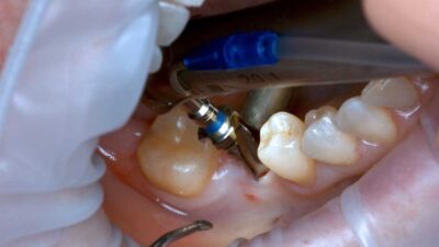
Following a healing period (typically 3-6 months) to allow for osseointegration, a second-stage surgery is performed to expose the implant abutment. This involves:
- Soft Tissue Management: A full-thickness mucoperiosteal flap is elevated to expose the implant. Careful soft tissue manipulation is essential to avoid damaging the peri-implant mucosa.
- Abutment Connection: The abutment is connected to the implant body. The choice of abutment depends on the prosthetic design and aesthetic requirements.
- Healing Cap Placement: A healing cap is placed on the abutment to shape the soft tissue contours.
7. Prosthetic Considerations
Prosthetic design should prioritize function, aesthetics, and long-term stability. Various prosthetic options exist, including:
- Single Crowns: Suitable for replacing single missing teeth.
- Fixed Partial Dentures (FPDs): Suitable for replacing multiple adjacent missing teeth.
- Overdentures: Suitable for replacing an entire arch of missing teeth.
- Implant-Supported Removable Partial Dentures (ISRPDs): Provide improved retention and stability compared to conventional RPDs.
8. Potential Complications & Management
Potential complications associated with two-stage implant placement include:
- Peri-implantitis: Inflammation of the soft tissues surrounding the implant. Management involves debridement, antibiotic therapy, and potentially surgical intervention.
- Implant Failure: Lack of osseointegration. Management involves implant removal and potential bone grafting.
- Soft Tissue Complications: Recession, inflammation, or hyperplasia. Management involves soft tissue grafting, mucogingival surgery, or crown lengthening.
- Nerve Damage: Can occur during implant placement in the posterior mandible. Prevention involves careful surgical planning and nerve localization techniques.
- Sinus Complications: Can occur during sinus lift procedures. Management involves antibiotic therapy and potentially sinus surgery.
- Mechanical Complications: Fracture of the implant, abutment, or restoration. Management involves replacement of the damaged component.
9. Conclusion
Two-stage implant placement remains a reliable and predictable technique for restoring partially edentate arches. Successful treatment requires meticulous patient selection, comprehensive diagnostics, precise surgical planning, and careful prosthetic design. Understanding the principles outlined in this review is crucial for optimizing treatment outcomes and providing long-term stability and function for patients with partial secondary edentulism. Advancements in guided implant surgery, bone grafting techniques, and prosthetic materials continue to improve the success rates and aesthetic outcomes of implant-supported rehabilitation.
Bibliography:
- Branemark, P. I., et al. (1969). Osseointegration. Clinical results and scientific background. Journal of Periodontology, 40(8), 626-647. (Original work describing osseointegration)
- Carlsson, L. E., & Albrektsson, T. (1994). Long-term results of fixed partial dentures supported by osseointegrated implants. International Journal of Oral & Maxillofacial Implants, 9(3), 357-364. (Early long-term study of implant-supported FPDs)
- Esposito, M., et al. (2009). Dental implant surfaces: a review of current knowledge and future perspectives. International Journal of Oral & Maxillofacial Implants, 24(Suppl), 179-194. (Review of implant surface characteristics)
- **Misch, C. E. (2008). *Dental implantology.* Elsevier Health Sciences. (Comprehensive textbook on implant dentistry)
- Simons, E. M., et al. (2017). The long-term survival and success of single-tooth implants in the aesthetic zone: a systematic review. International Journal of Oral & Maxillofacial Implants, 32(2), 343-360. (Review of long-term success of single implants)
- **Team EW. (2000). A systematic review of the effects of implant surface characteristics on osseointegration. *International Journal of Oral & Maxillofacial Implants*, *15*(Suppl), 50–60.
- Albrektsson, T., & Branemark, P. I. (1981). Principles of osseointegration. Quintessence International, 12(1), 39–54.
- Papaspyridakos, E., et al. (2010). Immediate or early implant loading in the aesthetic zone: a systematic review. International Journal of Periodontics & Restorative Dentistry, 30(4), 345-355. (Review of immediate loading)
- Vidi, A., et al. (2008). Sinus augmentation with bone grafts: a systematic review. Clinical Oral Implants Research, 19(1), 34-44. (Review of sinus lift procedures)
- Chen, S. T., et al. (2014). Guided tissue regeneration for peri-implantitis defects: a systematic review. International Journal of Oral & Maxillofacial Implants, 29(5), 925-935. (Review of peri-implantitis treatment)